What is cancer of the renal pelvis or ureter?
Cancer of the renal pelvis or ureter starts in the cells of the renal pelvis (a
hollow part of each
Cells in the renal pelvis or ureter sometimes change and no longer grow or behave
normally. In some cases, changes to these cells can cause cancer. Most often, cancer
starts in urothelial cells that line the inside of the renal pelvis or ureter. This
type of cancer is called urothelial carcinoma (also called transitional cell
carcinoma). It makes up about 90% of all upper
Rare types of renal pelvis and ureter cancer can also develop. These include squamous cell carcinoma and adenocarcinoma of the renal pelvis or ureter.
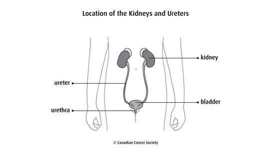
The renal pelvis and ureter
The renal pelvis and ureter are part of the
Urine (pee) is your body’s liquid waste and is made by the kidneys. It collects in the renal pelvis then travels along the ureters to the bladder where it is stored. When the bladder is full of urine, it passes out of the body through the urethra.
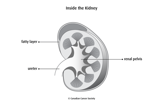
Layers of the renal pelvis and ureter
The walls of each renal pelvis and ureter are made up of 3 main layers of tissue.
The urothelium is the inner lining of each renal pelvis and ureter. It continues as the lining for the bladder and urethra. It is made up of urothelial cells (also called transitional cells), which can stretch and change shape as urine flows. The urothelium is also called the transitional epithelium.
The
lamina propria
is a thin layer of connective tissue that surrounds the urothelium of each
renal pelvis and ureter. It contains blood vessels, nerves and
The muscularis propria is a thick, outer muscle layer of each renal pelvis and ureter. It is made up of muscle that works automatically without you thinking about it (called smooth muscle). It pushes urine from the kidney down to the bladder.
Other tissues that surround the kidneys and ureters are:
- adventitia – loose connective tissue that covers the kidneys and ureters
- fat – a layer of fat that surrounds the renal pelvis, kidney and ureter
Cancerous tumours of the renal pelvis and ureter
Your trusted source for accurate cancer information
With support from readers like you, we can continue to provide the highest quality cancer information for over 100 types of cancer.
We’re here to ensure easy access to accurate cancer information for you and the millions of people who visit this website every year. But we can’t do it alone.
Every donation helps fund reliable cancer information, compassionate support services and the most promising research. Please give today because every contribution counts. Thank you.