The eyes
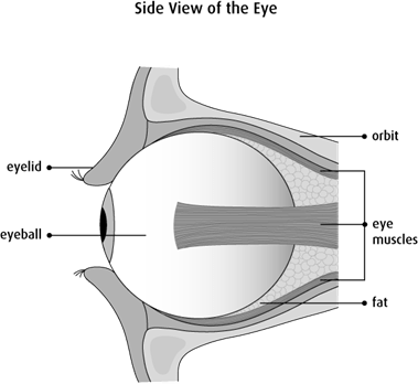
The eye is the organ of sight (vision). Only a small area of the eyeball can be seen because most of it is surrounded by the bones of the skull that make up the eye socket (orbit).
The eyeball
The main part of the eye is the eyeball. Each eye is shaped like a ball and its full size is about 2.5 cm (1 inch) in diameter. The inside of the eye contains a clear, jelly-like fluid that helps support the eye and maintain its shape. In the front of the eye this fluid is more watery and called aqueous humour. The fluid in the back of the eye is called vitreous humour.
The wall of the eye
The wall of the eye is the outer part of the eye that surrounds the ball of jelly-like fluid. It is made up of 3 layers called tunics. The wall of the eye contains many of the main parts of the eye.
- outer layer – made up of the sclera and cornea (called the fibrous tunic)
- middle layer – made up of the uvea (called the vascular tunic)
- inner layer – made up of the retina (called the neural tunic)
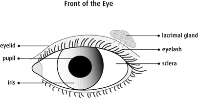
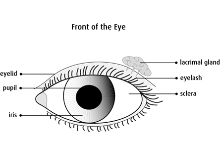
Sclera
The sclera is the white of the eye. It’s made up of tough connective tissue and covers most of the eyeball. The sclera is the protective covering of the eyeball. Muscles that control the movement of the eye attach to the sclera.
Cornea
The cornea is the clear, dome-shaped covering at the front of the eye. The cornea covers the pupil and the iris. It doesn’t have any blood vessels so cells in the cornea get oxygen from tears that cover the surface and the underlying aqueous humour. The lens is the main part that focuses light onto the retina but the cornea also plays a role in bending (refracting) light that enters the eye.
Uvea
The uvea is divided into 3 main parts – the iris, choroid and ciliary body. The uvea contains many blood vessels, lymph vessels and the inner muscles of the eye.
Iris
The iris is the thin, muscular coloured part of the eye. It is located between the cornea and the lens. The muscles of the iris change the size of the pupil (the small, black, central area of the eye) to control the amount of light that enters the eye. The iris has melanocytes, the cells that make a pigment called melanin. The amount of melanin in the iris is what gives the eye its colour.
Choroid
The choroid is a thin layer of tissue that lies between the sclera and retina. It contains many tiny blood vessels that supply oxygen and nutrients to the retina. The choroid contains many melanocytes. The melanocytes in the choroid absorb light to help lessen light reflection in the eye.
Ciliary body
The ciliary body is a muscular ring of tissue at the junction of the iris and the choroid. Muscle fibres in the ciliary body help the eye to focus on near or far objects by changing the shape of the lens. The ciliary body also has cells that make aqueous humour, the jelly-like fluid in the front of the eye between the cornea and lens.
Retina
The innermost layer of the wall of the eye is made up of the retina (also called the neural tunic). The retina has a layer of cells that absorb light that make up the pigmented layer. The retina also has a layer of nerve cells (neurons) that make up the neural retina. Some of the neurons of the neural retina are specialized cells called photoreceptors. There are 2 types of photoreceptors – rods and cones. Rods and cones are sensitive to light and work together like a camera to capture information about what we see.
Other neurons in the neural retina process some of the information about what we see and send it to the optic nerve. The optic nerve then sends this information to the brain to finish processing.
Lens
The lens is a transparent disc-shaped structure in the inner part of the eye. It lies directly behind the cornea and iris. The lens changes its shape to allow the eye to focus on near or far objects. Light rays pass through the lens and are focused on the retina to create images of objects at different distances from the body.
Accessory parts of the eye
The accessory (adnexal) parts of the eye include the eyelids, conjunctiva and lacrimal (tear) glands. They protect, lubricate and support the eyeball.
Eyelids
The eyelid is a fold of skin that covers and protects the eye. Muscles around the eye raise and close the eyelid. The eyelid has sebaceous glands that make an oily secretion that prevents the watery film on the eye from evaporating and the eyelid from sticking together. The eyelid works like a windshield wiper that helps to lubricate the eye and keep the surface of the eye free of dust and other debris. The eyelashes grow from the edges of the eyelid. They also help protect the eye from dust and debris.
Conjunctiva
The conjunctiva is a clear
Lacrimal gland and tears
The lacrimal gland (also called the tear gland) is located at the upper, outer corner of the eye. It secretes a watery fluid that makes up tears. Small ducts drain tears from the lacrimal gland through very tiny openings inside the inner corner of the eyelid. When the eye blinks, the tears are swept across the surface of the eye. Tears lubricate the conjunctiva covering the surface of the eye and inner eyelid. Tears also remove dust and debris from the eye and help to prevent infection.
Eye socket
The eye socket (orbit) is a bowl-shaped area made up of bone formed from the skull. It contains the eyeball, muscles, lacrimal gland, nerves, fat and connective tissues. The bone and tissues surrounding the eyeball help to cushion and protect it. Eye muscles allow the eyeball to move in different directions. These small muscles attach to the sclera near the front of the eye and to the bones of the orbit at the back.
Function of the eye
The eye and brain work together to allow us to see. The main function of the eye is to collect light and information about what we see. This information is sent to the brain through the optic nerve. The brain then turns information into a visual image or picture for us to see. If we lose the vision in one eye, we can still see most of what we could see before.