The lungs
The lungs are part of your
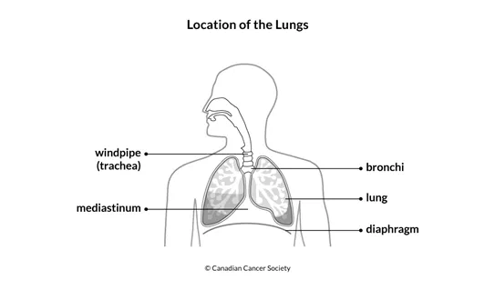
The windpipe (trachea) is a tube-shaped airway that leads to the lungs through the neck and chest. The windpipe further divides into 2 tubes that are the main airway to each lung. These airway tubes are called bronchi and they branch out into smaller and smaller tubes.
The lungs are separated by the
The lungs are surrounded by the chest wall. The chest wall is made up of the ribs and the muscles between the ribs.
Structure
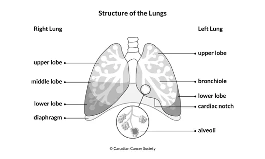
Each lung is divided into lobes (sections):
- The right lung has 3 lobes and is slightly larger than the left lung.
- The left lung has 2 lobes. The heart sits in the cardiac notch, which is a groove in the lower lobe.
Parts of the lung
The 2 main bronchi divide or branch into smaller bronchi, tubes which have small glands and cartilage in their walls. These smaller bronchi eventually divide into even smaller tubes called bronchioles, which have no glands or cartilage. At the end of the bronchioles are millions of tiny air sacs called alveoli. Surrounding the alveoli are very tiny blood vessels (called capillaries).
The bronchi are lined with cells that have very fine hair like projections called cilia.
Each lung is covered by a thin membrane called the pleura. The pleura also lines the inner side of the rib cage. It protects and cushions the lungs and produces a fluid that acts like a lubricant so the lungs can move smoothly in the chest cavity. The pleura is made up of 2 layers:
- inner (visceral) pleura – the layer next to the lung
- outer (parietal) pleura – the layer that lines the chest wall
The area between the 2 layers of the pleura is called the pleural space.
The lymphatic system of the lung
Different groups of lymph nodes, which are part of the
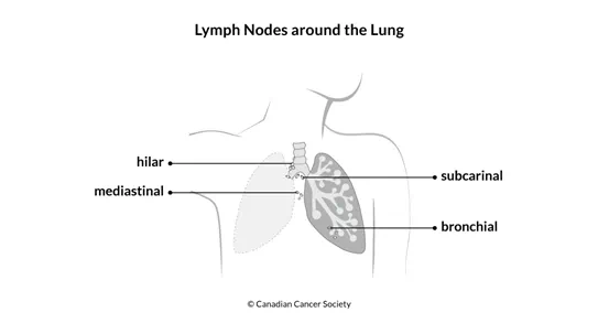
- bronchial nodes – lymph nodes in the lung around the ends of the bronchi
- hilar nodes – lymph nodes in the area where the windpipe divides into the main bronchi
- mediastinal nodes – lymph nodes along the windpipe in between the 2 lungs
- subcarinal nodes – lymph nodes just below the windpipe where it divides into the main bronchi
What the lungs do
The main functions of the lungs are to transfer oxygen from the air to the blood and to release carbon dioxide from the blood to the air.
- Air enters the mouth or nose and travels through the windpipe, bronchi and bronchioles to the alveoli. The exchange of oxygen and carbon dioxide happens in the alveoli.
- The alveoli absorb oxygen from the air and pass it into the blood, which circulates the oxygen around the body.
- Carbon dioxide, which is a waste product of the body's cells, passes from the blood into the alveoli and is breathed out.
- The lungs produce a mixture of fats and proteins called lung or pulmonary surfactant. The surfactant coats the surfaces of the alveoli, making it easier for them to expand and deflate with each breath.
The lungs also help protect the body from harmful substances in the air, such as smoke, pollution, bacteria and viruses. These substances can pass through the nose and become trapped in the lungs. The lungs produce a thick, slippery fluid (mucus), which can trap and partly destroy these substances from the air. The cilia move rapidly to push the mucus up through the bronchi, where it is removed by coughing or swallowing.