What is esophageal cancer?
Esophageal cancer starts in the cells of the esophagus. A cancerous (malignant) tumour is a group of cancer cells that can grow into and destroy nearby tissue. It can also spread (metastasize) to other parts of the body.
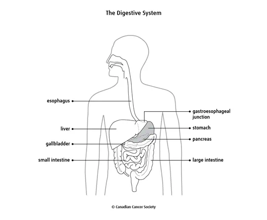
The esophagus is part of the digestive system. It is part of the gastrointestinal (GI) tract, which is the tube that goes from the mouth to the anus. The GI tract includes the esophagus, stomach, small intestine, large intestine and other organs.
Types of esophageal cancer
Cells in the esophagus sometimes change and no longer grow or behave normally. These changes may lead to non-cancerous (benign) conditions such as esophageal webs and rings. They can also lead to non-cancerous tumours such as leiomyomas
Changes to cells of the esophagus can also cause precancerous conditions. This means that the abnormal cells are not yet cancer, but there is a chance that they may become cancer if they aren't treated. The most common precancerous condition of the esophagus is Barrett's esophagus.
But in some cases, changes to the cells that line the esophagus can cause esophageal cancer. Most often, esophageal cancer starts in the glandular cells which make mucus. This type of cancer is called adenocarcinoma of the esophagus. Cancer can also start in the flat, thin cells called squamous cells. This type of cancer is called squamous cell carcinoma of the esophagus.
Rare types of esophageal cancer can also develop. These include gastrointestinal stromal tumour (GIST), leiomyosarcoma and neuroendocrine tumours (NETs).
The esophagus
The esophagus is a hollow, muscular tube that allows food to pass from the mouth to the stomach. It is 25–33 cm long and less than 2.5 cm cross at its narrowest part. The esophagus begins in the throat, or pharynx, and connects to the stomach. In the throat, it lies behind the windpipe, or trachea. In the chest cavity, it is in front of the backbone, or spine.
There are a number of
Layers of the esophagus
The esophagus has 3 layers of tissue and a covering.
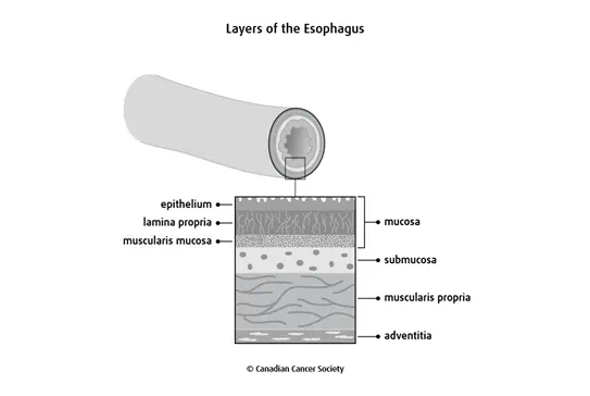
The mucosa is the inner lining of the esophagus. It is made up of: ·
- a thin layer of squamous cells (called the epithelium)
- a layer of connective tissue (called the lamina propria)
- a thin layer of muscle (called the muscularis mucosa)
The submucosa is a layer of connective tissue that surrounds the mucosa layer. It contains mucous glands, blood vessels, nerves and lymphatic tissue.
The muscularis propria lies outside of the submucosa layer. It has an inner ring of circular muscle fibres and an outer ring of long muscle fibres that surround the wall of the esophagus.
The adventitia is a layer of connective tissue that loosely supports and covers the outside of the esophagus in the neck and chest.
Function
Although the esophagus is part of the digestive system, it doesn't actually play a role in digesting food. But it is still an important organ in the GI tract.
When you swallow, the muscular walls of the esophagus tighten, or contract, to push food and fluids from the mouth into the stomach. Glands in the submucosa layer of the esophagus make mucus, which helps keep the inside of the esophagus moist and gives lubrication for swallowing.
The upper esophageal sphincter is a ring of muscle fibres. It detects food and liquids in the back of the throat. When you swallow this sphincter closes, allowing food to pass from the mouth into the esophagus and preventing food from going into the windpipe (trachea).
The wall of the esophagus includes 2 layers of strong muscles. These muscles tighten, or contract, to help push food along until it reaches the stomach. The series of contractions that move food through the esophagus is called peristalsis.
There is another ring of muscle fibres at the bottom of the esophagus. These muscle fibres form the lower esophageal sphincter (also known as the cardiac sphincter). This sphincter prevents food from moving up out of the stomach. The area where the esophagus joins the stomach is called the gastroesophageal (GE) junction. The GE junction is close to the diaphragm, the thin muscle that separates the chest cavity from the abdomen.
At the GE junction, the lower esophageal sphincter acts like a valve to control the passage of food into the stomach. When it opens, the valve lets food into the stomach. When closed, it prevents the stomach contents from going back into the esophagus.