The breasts
Women and men both have breasts, but women have more breast tissue than men. The breasts contain mammary glands. Only the mammary glands in women can make milk to feed a baby. For this reason, the breasts are accessory organs of the female reproductive system.
The breasts are divided by an invisible line running up and down and right to left through the nipple. Each of these 4 regions is called a quadrant. Most breast cancers develop in the upper outer quadrant of the breast, closest to the armpit. This is because this area has a lot of glandular tissue.
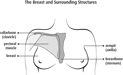
Structure
Each breast lies over a large muscle (called the pectoralis major muscle) on the chest. The breast covers a fairly large area. It goes from just below the collarbone (called the clavicle) to the armpit (called the axilla) and across to the breastbone (called the sternum).
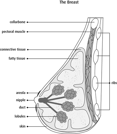
Parts of the breast
The breast is made up of fat, connective tissue, glands and ducts.
Ligaments are dense bands of connective tissue that support the breast. They run from the skin through the breast and attach to muscles on the chest.
Lobules are the groups of glands that make milk. There are 15–25 lobules in each breast. The glands make milk when they are stimulated by hormones in a woman’s body during pregnancy.
Ducts are tubes that carry milk from the lobules to the nipple.
The nipple is the area at the centre of the areola with an opening to release milk. The nipples contain muscle fibres. When these muscle fibres contract, the nipple becomes erect, or pointed outward.
The areola is the pink or brown, circular area around the nipple on the surface of the breast. It contains small glands that release, or secrete, an oily substance that acts as a lubricant for the nipple and areola.
The lymphatic system of the breast
The breast has many blood vessels and lymph vessels. Lymph vessels are thin tubes similar
to blood vessels. They collect and move lymph fluid away from the breast into small
bean-shaped masses of lymphatic tissue (called lymph nodes) around the breast. The lymph
vessels and lymph nodes are part of the
There are several groups of lymph nodes that drain each breast. These groups of lymph nodes are found on both sides of the body.
The supraclavicular lymph nodes are above the collarbone.
The infraclavicular, or subclavicular, lymph nodes are below the collarbone.
The internal mammary lymph nodes are inside the chest around the breastbone (called sternum).

The axillary lymph nodes are under the arm (called the axilla). There are about 30–50 lymph nodes in the axilla. They are divided into 3 levels based on how close they are to the large muscle of the chest (called the pectoralis major). When breast cancer spreads, it usually spreads to level I lymph nodes, then to level II and then to level III.
- Level I, or low axilla, are along the outer border of the muscle under the pectoralis major (called the pectoralis minor)
- Level II, or mid axilla, are beneath the pectoralis minor.
- Level III, or high axilla, are along the inner border of the pectoralis minor.

Breast development in women
Breasts start to grow during puberty, which usually takes place between 10 and 12 years of age. At this time, the breasts respond to hormonal changes in the body. These changes include higher estrogen and progesterone levels in the body. The breasts start to build up fat and the ducts begin to grow and branch. The glands of the breast also start to develop during puberty. The breast skin stretches as the breasts grow, creating a rounded appearance.
Young women tend to have denser breasts with more gland tissue than older women. As a woman ages, much of the gland and duct tissues are replaced with fat. This makes the breasts less dense. Ligaments also lose their elasticity as a woman ages, which causes the breasts to change shape and lose some of their fullness.
Women’s breasts vary in size and shape. The size depends on the amount of fat in the breast. A woman’s breasts are rarely the same size. One breast is usually slightly larger or smaller, higher or lower or shaped differently than the other.
Breast changes during menstruation and menopause
Estrogen and progesterone cause changes to the breast tissues each menstrual cycle. In the first part of the menstrual cycle, estrogen stimulates the milk ducts to make them grow. Progesterone takes over in the second part of a woman’s menstrual cycle. It stimulates the glands in the lobules so they are ready to make milk if a woman becomes pregnant.
After menopause, the ovaries don’t make estrogen and progesterone so breasts don’t go through these monthly changes.
Breast changes during pregnancy and lactation
The main function of a woman’s breasts is to make, store and release milk to feed a baby. After giving birth, hormones in a woman’s body stimulate the glands in the lobules throughout the breast to make milk. The ducts carry the milk to the nipple. Milk passes from the nipple to the baby during breast-feeding.
The breasts of women who aren’t pregnant or breast-feeding contain mostly fat and ducts. There is a small amount of gland tissue, but the breast glands don’t develop fully until the 6th month of pregnancy in most women.
The breasts of pregnant and breast-feeding women contain mostly gland tissue that makes milk. This is why a woman’s breasts tend to get bigger during pregnancy and when a woman is breast-feeding.
Hormones and the breast
Estrogen is the main female hormone. It influences female sexual characteristics, such as breast development, and it is necessary for reproduction. Most of the estrogen in a woman’s body is made by the ovaries, but a small amount is made by the adrenal glands.
Progesterone is the other female sex hormone made in the ovaries. Its role is to prepare the uterus, or womb, for pregnancy during each menstrual cycle. Progesterone also plays a role in completing the development of the mammary glands during pregnancy so they can make milk after childbirth.