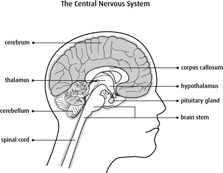
The brain and spinal cord
The brain is a complex organ made up of specialized nerve and supportive tissues. It’s surrounded by many bones that together form the skull. The part of the skull where the brain sits is called the cranium. The base, or lower part, of the brain is connected to the spinal cord. Together, the brain and spinal cord are known as the central nervous system (CNS). Many nerves send electrical signals to and from the brain and spinal cord.
Structure and function of the brain
The brain is the body’s control centre. It constantly receives and interprets nerve signals from the body and sends new signals based on this information. Different parts of the brain control movement, speech, emotions, consciousness and internal body functions, such as heart rate, breathing and body temperature.
The brain has 3 main parts: cerebrum, cerebellum and brain stem.
Types of cells in the brain
The brain is made up of 2 main types of cells:
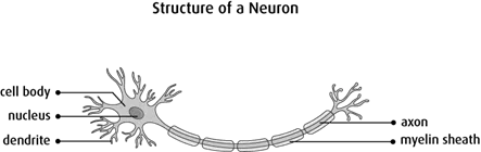
Nerve cells (neurons) are cells that carry the electrical signals that make the nervous system work. They cannot be replaced or repaired if they are damaged. They are the longest cells in the body.
Glial cells (neuroglial cells) are cells that support, feed and protect the nerve cells. The different types of glial cells are:
- astrocytes
- oligodendrocytes
- ependymal cells
- microglial cells
Cerebrum
The cerebrum is the largest part of the brain. It is divided into 2 halves called the left and right cerebral hemispheres. The 2 hemispheres are connected by a bridge of nerve fibres called the corpus callosum.
The right half of the cerebrum (right hemisphere) controls the left side of the body. The left half of the cerebrum (left hemisphere) controls the right side of the body.
The cerebral cortex is the outer, folded part of the brain. It is also called the grey matter. The cerebral cortex is mostly made up of the cell bodies and dendrites of nerve cells (neurons). Cell bodies contain the nucleus and other main parts of the cell. Dendrites are the short branching fibres that receive signals from other nerve cells. The inner part of the cerebrum is called the white matter. It is mostly made up of the long fibres of a nerve cell (called axons) that send signals to and from the brain to the rest of the body. The fatty coating that surrounds axons (called myelin) gives this part of the brain a whitish appearance.
Each hemisphere is divided into 4 sections called lobes. These include the frontal, parietal, temporal and occipital lobes.
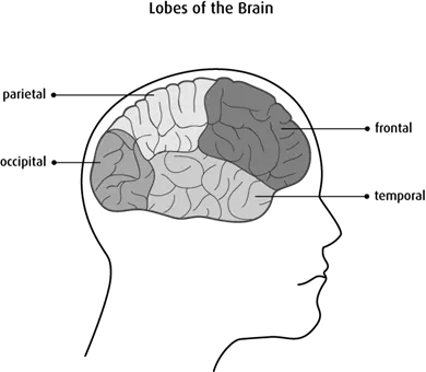
Each lobe has different functions:
The frontal lobe controls movement, speech, behaviour, memory, emotions and intellectual functions, such as thought processes, reasoning, problem solving, decision-making and planning.
The parietal lobe controls sensations, such as touch, pressure, pain and temperature. It also controls the understanding of size, shape and direction (called spatial orientation).
The temporal lobe controls hearing, memory and emotions. The dominant (left side in most right-handed people) temporal lobe also controls speech.
The occipital lobe controls vision.
Cerebellum
The cerebellum is located under the cerebrum at the back of the brain. It is divided into 2 parts or hemispheres and also has grey and white matter.
The cerebellum is responsible for:
- movement
- posture
- balance
- reflexes
- complex actions (walking, talking)
- collecting sensory information from the body
Brain stem
The brain stem is a bundle of nerve tissue at the base of the brain. It connects the cerebrum and cerebellum to the spinal cord.
The brain stem has 3 areas:
- midbrain (also called the mesencephalon)
- pons
- medulla oblongata
The brain stem sends information to and from the other parts of the brain to the rest of the body and controls:
- breathing
- body temperature
- blood pressure
- heart rate
- hunger and thirst
- digestion of food
Cerebrospinal fluid (CSF)
The cerebrospinal fluid (CSF) is a clear, watery liquid that surrounds, cushions and protects the brain and spinal cord. The CSF also carries nutrients in the blood to (and removes waste products from) the brain. It circulates through chambers called ventricles and over the surface of the brain and spinal cord.
Meninges
The brain and spinal cord are covered and protected by 3 layers of tissue (membranes) called the meninges:
- dura mater – thickest outer membrane
- arachnoid layer – middle, thin membrane
- pia mater – inner, thin membrane
The CSF flows in the space between the arachnoid layer and the pia mater. This space is called the subarachnoid space.
Corpus callosum
The corpus callosum is a bundle of nerve fibres that allows communication between the 2 cerebral hemispheres. It is the largest fibre bundle in the brain.
Thalamus
The thalamus is a structure in the middle of the brain that has 2 lobes or sections. It acts as a relay station for almost all information that comes and goes between the brain and the rest of the nervous system in the body.
Hypothalamus
The hypothalamus is a small structure in the middle of the brain below the thalamus. It plays a part in controlling body temperature,
Pituitary gland
The pituitary gland is a small, pea-sized organ in the centre of the brain. It is attached to the hypothalamus and makes a number of different hormones that affect other glands of the body’s
Pineal gland
The pineal gland is a very small gland in the third ventricle of the brain. It produces the hormone melatonin, which influences sleeping and waking patterns and sexual development.
Choroid plexus
The choroid plexus is a small organ in the ventricles that makes CSF.
Cranial nerves
There are 12 pairs of cranial nerves that perform specific functions in the head and neck, including giving us our sense of smell, sight (vision), hearing, taste, speech, feeling in the face and movement of the muscles in the face, eyes and tongue. One pair of nerves starts in specialized cells in the roof of the nose and another pair starts in the retina of the eye. The other 10 pairs start in the brain stem.
Blood-brain barrier (BBB)
The blood-brain barrier (BBB) is a specialized system of cells lining blood vessels in the brain. The BBB prevents most substances in the blood from passing into the brain and helps maintain a constant environment so the nerve cells in the brain can work properly.
The BBB is made up of very small blood vessels (capillaries) that are lined with thin, flat endothelial cells. In other parts of the body, endothelial cells have small spaces between them that allow substances to move in and out of the capillary so they can reach other cells and tissues. In the brain, the endothelial cells are packed tightly together so substances cannot pass out of the bloodstream into the brain.
Structure and function of the spine
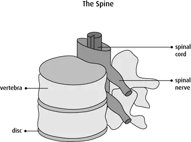
The spine is made up of 26 bones divided into 5 sections. These bones surround and protect the spinal cord. This includes 24 vertebrae (divided into cervical, thoracic and lumbar regions), the sacrum and the coccyx.
Cervical region – These are 7 vertebrae at the top of the spine that run from the base of the skull to the lowest part of the neck.
Thoracic region – These are 12 vertebrae that run from the shoulders to the middle of the back.
Lumbar region – These are 5 vertebrae that run from the middle of the back to the hips.
Sacrum – This is a large section of fused vertebrae at the base of the spine.
Coccyx (tail bone) – This is a small, thin section of fused vertebrae at the end of the spine.
Between the vertebrae are the discs (intervertebral discs).
Disc – A layer of cartilage found between the vertebrae. Discs cushion and protect the vertebrae and spinal cord.
Spinal cord
The spinal cord is a thick column of nerves surrounded by vertebrae that runs from the brain stem to the lumbar region of the spine. Like the brain, the spinal cord has both grey and white matter. The spinal cord sends information between the brain and most of the body through the spinal nerves.
Spinal nerves
Pairs of spinal nerves exit the vertebrae along the length of the spinal cord. At the lumbar region, the spinal cord branches into a group of spinal nerves that exit the lumbar vertebrae and sacrum. The spinal nerves control body functions like movement, bladder and bowel control and breathing. The spinal nerves are numbered after nearby vertebrae.