The bladder

The bladder is part of the urinary system. It is a hollow organ in the pelvis that stores urine (pee) before it leaves the body. The wall of the bladder is stretchy so it gets bigger (expands) as it fills with urine and gets smaller (contracts) as it empties.

Diagram of the location of the bladder

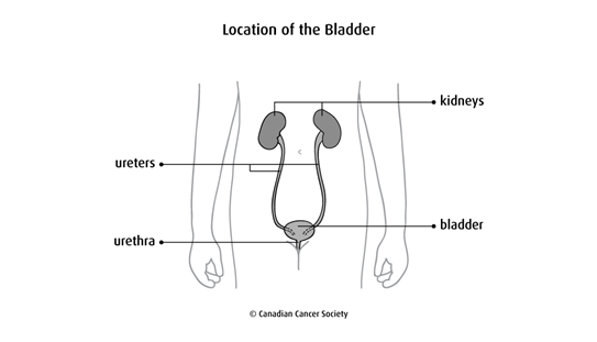
How the bladder works

Urine is made by the kidneys and travels to the bladder along 2 long tubes called ureters. When the bladder is full of urine, muscles in the bladder wall tighten and push the urine out of the bladder through the urethra where it leaves the body.

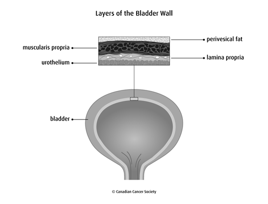
Layers of the bladder wall

The bladder wall is made up of 3 main layers.

The urothelium is the inner lining of the bladder. It is made up of urothelial cells (also called transitional cells). The urothelium is also called the transitional epithelium.

The lamina propria (also called the submucosa) is a thin layer of connective tissue that surrounds the urothelium. It contains blood vessels, nerves and glands.

The muscularis propria is the thick, outer muscle layer of the bladder. It is made up of 3 layers of muscle that work automatically without you thinking about it (called smooth muscle).

Diagram of the layers of the bladder wall

Other tissue that separates the bladder from other organs are:

  • serosa – a thin membrane that covers the top part of the bladder
  • adventitia – loose connective tissue that covers areas of the bladder where there is no serosa
  • perivesical fat – a layer of fat surrounding the bladder

Expert review and references

  • Al-Ahmadie H, Lin O, Reuter VE . Pathology and cytology of tumors of the urinary tract. Scardino PT, Lineham WM, Zelefsky MJ, Vogelzang NJ (eds.). Comprehensive Textbook of Genitourinary Oncology. 4th ed. Philadelphia: Wolters Kluwer/Lippincott Williams & Wilkins; 2011: 16:295-316.
  • American Cancer Society . About Bladder Cancer . 2016 : https://www.cancer.org/.
  • American Society of Clinical Oncology . Bladder Cancer . 2017 : https://www.cancer.net/.
  • Martini FH, Timmons MJ, Tallitsch RB. Human Anatomy. 7th ed. San Francisco: Pearson Benjamin Cummings; 2012.
  • National Cancer Institute . Bladder Cancer Treatment (PDQ®) Health Professional Version . 2018 : https://www.cancer.gov/.
  • Surveillance Epidemiology and End Results (SEER), National Cancer Institute . SEER Training Modules: Anatomy of the Bladder . Bethesda, MD : National Cancer Institute ; https://training.seer.cancer.gov/index.html.

Medical disclaimer

The information that the Canadian Cancer Society provides does not replace your relationship with your doctor. The information is for your general use, so be sure to talk to a qualified healthcare professional before making medical decisions or if you have questions about your health.

We do our best to make sure that the information we provide is accurate and reliable but cannot guarantee that it is error-free or complete.

The Canadian Cancer Society is not responsible for the quality of the information or services provided by other organizations and mentioned on cancer.ca, nor do we endorse any service, product, treatment or therapy.


1-888-939-3333 | cancer.ca | © 2024 Canadian Cancer Society