The small intestine
The small intestine is also called the small bowel. It is a hollow, tube-like organ that
is connected to the stomach on one end and the large intestine on the other. The small
intestine is the longest part of the
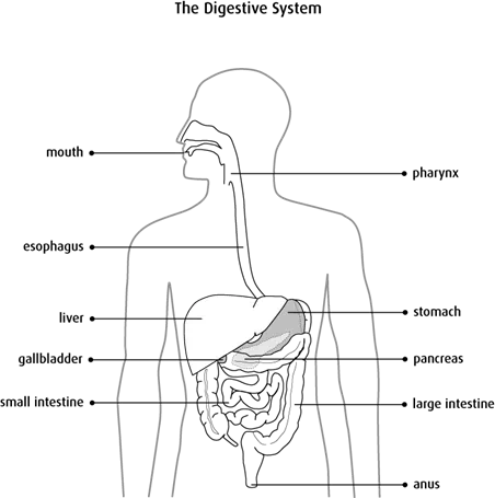
The GI tract is a tube that goes from the mouth to the anus. The esophagus carries food from the mouth and throat to the stomach. The stomach receives food from the esophagus, starts the digestive process and empties partly digested food into the small intestine. The small intestine continues digestion and absorbs nutrients. The large intestine absorbs water from partly digested food, forms it into stool and stores it until it is passed out of the body with a bowel movement.
Several other organs of the digestive system help to digest food, including the liver, gallbladder and pancreas.
Structure
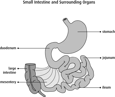
The small intestine is about 4.75–6 m (15–20 ft) long. It has an average diameter of 2.5 cm (1 in). It is looped and coiled and fills up much of the abdominal cavity (the space in the abdomen that contains the intestines and other organs). The small intestine is made up of the duodenum, jejunum and ileum. It is covered by mesentery.
Duodenum
The duodenum connects the stomach to the small intestine. Most digestive
Jejunum
The jejunum is the middle part of the small intestine, between the duodenum and ileum. Most digestion and nutrient absorption takes place in the jejunum.
Ileum
The ileum is the last and longest part of the small intestine. The ileum absorbs nutrients from digested food and empties the waste into the large intestine.
Mesentery
The mesentery is a membrane that suspends the small intestine from the back of the abdominal wall. It supports the blood vessels, nerves and lymph vessels that supply the small intestine. The small intestine is surrounded by the large intestine.
Layers of the small intestine wall
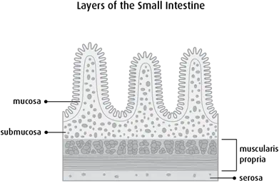
The small intestine is made up of the same 4 layers that make up most of the GI tract.
Mucosa
The mucosa is a mucous membrane. It is the innermost lining of the small intestine. It is made up of a:
- layer of
epithelial cells (called the epithelium) - layer of loose connective tissue (called the lamina propria)
- very thin layer of muscle (called the muscularis mucosa)
The inner surface of the mucosa has many finger-like projections called villi. The villi increase the surface area of the small intestine, which helps it absorb digested food.
Submucosa
The submucosa is a layer of connective tissue that surrounds the mucosa. It contains mucous glands, blood vessels, lymph vessels and nerves.
Muscularis propria
The muscularis propria lies outside the submucosa. It is a band of smooth muscle that helps move food through the small intestine.
Serosa
The serosa is the outermost layer that covers the small intestine. It is formed by the visceral layer of the peritoneum (layers of tissue that cover the outer surface of most organs in the abdomen). The mesentery is attached to the serosa.
Function
The main functions of the small intestine are to break down, or digest, food and to absorb nutrients, such as