What is gestational trophoblastic disease?
Gestational trophoblastic disease (GTD) includes several rare tumours that occur inside the uterus (womb) and start in the cells that form the placenta during pregnancy. Most gestational trophoblastic diseases are non-cancerous. It is estimated that the cancerous forms of GTD make up less than 1% of all women’s reproductive system cancers.
The uterus is part of a woman’s reproductive system. The uterus is the organ where a fetus (baby) develops and grows during pregnancy.
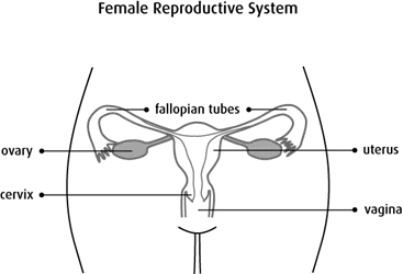
The function of the uterus is to receive a fertilized egg and protect the baby while it grows and develops. The uterus contracts to push the baby out of the body during birth.
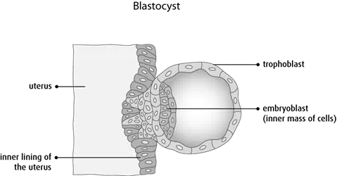
Early in pregnancy, the fertilized egg divides and grows to become a mass of cells called a blastocyst. The blastocyst normally attaches to the lining of the uterus a few days after conception. The blastocyst contains an early embryo and trophoblastic cells.
The embryocyst is the inner mass of cells that will become the embryo. The trophoblast is the outer layer of cells surrounding the embryo. It has many small projections (called chorionic villi) that grow into the lining of the uterus. The layers of the trophoblast form the tissue that will become the placenta and the membranes that surround the embryo. The placenta is attached to the lining of the uterus and provides a way for blood, oxygen and nutrients to move from the mother to the developing fetus.
In rare cases, a pregnancy does not progress normally and may form a hydatidiform mole (molar pregnancy). These pregnancies lack an embryo and have abnormal growth of trophoblastic tissue. The body can abort hydatidiform moles completely (spontaneous abortion or miscarriage). If they do not abort completely, the woman will need a procedure to completely remove the abnormal tissue from the uterus.
GTD can occur:
- during a pregnancy
- after a miscarriage or surgical removal of a pregnancy (abortion)
- after a tubal pregnancy (implantation of the fertilized egg within the fallopian tube)
- after a normal pregnancy